 |
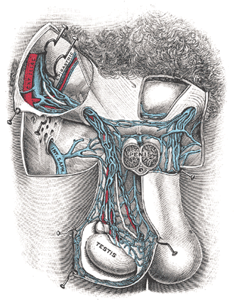
| FIG. 1143– The scrotum. On the left side the cavity of the tunica vaginalis has been opened; on the right side only the layers superficial to the Cremaster have been removed. (Testut.) |
|
(Organa Genitalia Virilia) The male genitals include the testes, the ductus deferentes, the vesiculæ seminales, the ejaculatory ducts, and the penis, together with the following accessory structures, viz., the prostate and the bulbourethral glands. |
| |
| 1. The Testes and their Coverings (Figs. 1143, 1144, 1145)—The testes are two glandular organs, which secrete the semen; they are suspended in the scrotum by the spermatic cords. At an early period of fetal life the testes are contained in the abdominal cavity, behind the peritoneum. Before birth they descend to the inguinal canal, along which they pass with the spermatic cord, and, emerging at the subcutaneous inguinal ring, they descend into the scrotum, becoming invested in their course by coverings derived from the serous, muscular, and fibrous layers of the abdominal parietes, as well as by the scrotum. |
| The coverings of the testes are, the |
| Skin | Scrotum. | Cremaster. |
| Dartos tunic | Infundibuliform fascia. |
| Intercrural fascia. | | Tunica vaginalis. | |
| |
| The Scrotum is a cutaneous pouch which contains the testes and parts of the spermatic cords. It is divided on its surface into two lateral portions by a ridge or raphé, which is continued forward to the under surface of the penis, and backward, along the middle line of the perineum to the anus. Of these two lateral portions the left hangs lower than the right, to correspond with the greater length of the left spermatic cord. Its external aspect varies under different circumstances: thus, under the influence of warmth, and in old and debilitated persons, it becomes elongated and flaccid; but, under the influence of cold, and in the young and robust, it is short, corrugated, and closely applied to the testes. |
| The scrotum consists of two layers, the integument and the dartos tunic. |
| The Integument is very thin, of a brownish color, and generally thrown into folds or rugæ. It is provided with sebaceous follicles, the secretion of which has a peculiar odor, and is beset with thinly scattered, crisp hairs, the roots of which are seen through the skin. |
| The Dartos Tunic (tunica dartos) is a thin layer of non-striped muscular fibers, continuous, around the base of the scrotum, with the two layers of the superficial fascia of the groin and the perineum; it sends inward a septum, which divides the scrotal pouch into two cavities for the testes, and extends between the raphé and the under surface of the penis, as far as its root. |
 |
| FIG. 1144– The scrotum. The penis has been turned upward, and the anterior wall of the scrotum has been removed. On the right side, the spermatic cord, the infundibuliform fascia, and the Cremaster muscle are displayed; on the left side, the infundibuliform fascia has been divided by a longitudinal incision passing along the front of the cord and the testicle, and a portion of the parietal layer of the tunica vaginalis has been removed to display the testicle and a portion of the head of the epididymis, which are covered by the visceral layer of the tunica vaginalis. (Toldt.) |
| |
| The dartos tunic is closely united to the skin externally, but connected with the subjacent parts by delicate areolar tissue, upon which it glides with the greatest facility. |
| The Intercrural Fascia (intercolumnar or external spermatic fascia) is a thin membrane, prolonged downward around the surface of the cord and testis (see page 411). It is separated from the dartos tunic by loose areolar tissue. |
| The Cremaster consists of scattered bundles of muscular fibers connected together into a continuous covering by intermediate areolar tissue (see page 414). |
| The Infundibuliform Fascia (tunica vaginalis communis [testis et funiculi spermatici]) is a thin layer, which loosely invests the cord; it is a continuation downward of the transversalis fascia (see page 418). |
| The Tunica Vaginalis is described with the testes. |
 |
| FIG. 1145– Transverse section through the left side of the scrotum and the left testis. The sac of the tunica vaginalis is represented in a distended condition. (Diagrammatic.) (Delépine.) |
| |
| |
| Vessels and Nerves.—The arteries supplying the coverings of the testes are: the superficial and deep external pudendal branches of the femoral, the superficial perineal branch of the internal pudendal, and the cremasteric branch from the inferior epigastric. The veins follow the course of the corresponding arteries. The lymphatics end in the inguinal lymph glands. The nerves are the ilioinguinal and lumboinguinal branches of the lumbar plexus, the two superficial perineal branches of the internal pudendal nerve, and the pudendal branch of the posterior femoral cutaneous nerve. |
| The Inguinal Canal (canalis inguinalis) is described on page 418. |
| The Spermatic Cord (funiculus spermaticus) (Fig. 1146) extends from the abdominal inguinal ring, where the structures of which it is composed converge, to the back part of the testis. In the abdominal wall the cord passes obliquely along the inguinal canal, lying at first beneath the Obliquus internus, and upon the fascia transversalis; but nearer the pubis, it rests upon the inguinal and lacunar ligaments, having the aponeurosis of the Obliquus externus in front of it, and the inguinal falx behind it. It then escapes at the subcutaneous ring, and descends nearly vertically into the scrotum. The left cord is rather longer than the right, consequently the left testis hangs somewhat lower than its fellow. |
| |
| Structure of the Spermatic Cord.—The spermatic cord is composed of arteries, veins, lymphatics, nerves, and the excretory duct of the testis. These structures are connected together by areolar tissue, and invested by the layers brought down by the testis in its descent. |
| The arteries of the cord are: the internal and external spermatics; and the artery to the ductus deferens. |
| The internal spermatic artery, a branch of the abdominal aorta, escapes from the abdomen at the abdominal inguinal ring, and accompanies the other constituents of the spermatic cord along the inguinal canal and through the subcutaneous inguinal ring into the scrotum. It then descends to the testis, and, becoming tortuous, divides into several branches, two or three of which accompany the ductus deferens and supply the epididymis, anastomosing with the artery of the ductus deferens: the others supply the substance of the testis. |
| The external spermatic artery is a branch of the inferior epigastric artery. It accompanies the spermatic cord and supplies the coverings of the cord, anastomosing with the internal spermatic artery. |
| The artery of the ductus deferens, a branch of the superior vesical, is a long, slender vessel, which accompanies the ductus deferens, ramifying upon its coats, and anastomosing with the internal spermatic artery near the testis. |
 |
| FIG. 1146– The spermatic cord in the inguinal canal. (Poirier and Charpy.) |
| |
| The spermatic veins (Fig. 1147) emerge from the back of the testis, and receive tributaries from the epididymis: they unite and form a convoluted plexus, the plexus pampiniformis, which forms the chief mass of the cord; the vessels composing this plexus are very numerous, and ascend along the cord in front of the ductus deferens; below the subcutaneous inguinal ring they unite to form three or four veins, which pass along the inguinal canal, and, entering the abdomen through the abdominal inguinal ring, coalesce to form two veins. These again unite to form a single vein, which opens on the right side into the inferior vena cava, at an acute angle, and on the left side into the left renal vein, at a right angle. |
| The lymphatic vessels are described on page 713. |
| The nerves are the spermatic plexus from the sympathetic, joined by filaments from the pelvic plexus which accompany the artery of the ductus deferens. |
| The scrotum forms an admirable covering for the protection of the testes. These bodies, lying suspended and loose in the cavity of the scrotum and surrounded by serous membrane, are capable of great mobility, and can therefore easily slip about within the scrotum and thus avoid injuries from blows or squeezes. The skin of the scrotum is very elastic and capable of great distension, and on account of the looseness and amount of subcutaneous tissue, the scrotum becomes greatly enlarged in cases of edema, to which this part is especially liable as a result of its dependent position. |
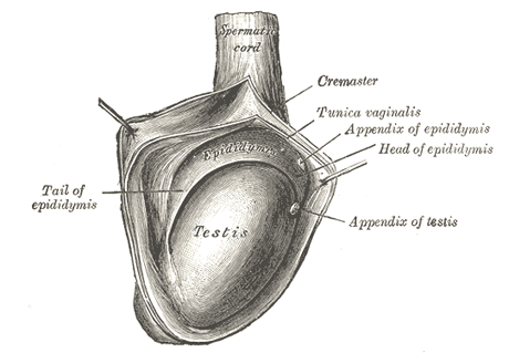
| The Testes are suspended in the scrotum by the spermatic cords, the left testis hanging somewhat lower than its fellow. The average dimensions of the testis are from 4 to 5 cm. in length, 2.5 cm. in breadth, and 3 cm. in the antero-posterior diameter; its weight varies from 10.5 to 14 gm. Each testis is of an oval form (Fig. 1148), compressed laterally, and having an oblique position in the scrotum; the upper extremity is directed forward and a little lateralward; the lower, backward and a little medialward; the anterior convex border looks forward and downward, the posterior or straight border, to which the cord is attached, backward and upward. |
| The anterior border and lateral surfaces, as well as both extremities of the organ, are convex, free, smooth, and invested by the visceral layer of the tunica vaginalis. The posterior border, to which the cord is attached, receives only a partial investment from that membrane. Lying upon the lateral edge of this posterior border is a long, narrow, fiattened body, named the epididymis. |
 |
| FIG. 1147– Spermatic veins. (Testut.) |
| |
 |
| FIG. 1148– The right testis, exposed by laying open the tunica vaginalis. |
| |
| The epididymis consists of a central portion or body; an upper enlarged extremity, the head (globus major); and a lower pointed extremity, the tail (globus minor), which is continuous with the ductus deferens, the duct of the testis. The head is intimately connected with the upper end of the testis by means of the efferent ductules of the gland; the tail is connected with the lower end by cellular tissue, and a reflection of the tunica vaginalis. The lateral surface, head and tail of the epididymis are free and covered by the serous membrane; the body is also completely invested by it, excepting along its posterior border; while between the body and the testis is a pouch, named the sinus of the epididymis (digital fossa). The epididymis is connected to the back of the testis by a fold of the serous membrane. |
| |
| Appendages of the Testis and Epididymis.—On the upper extremity of the testis, just beneath the head of the epididymis, is a minute oval, sessile body, the appendix of the testis (hydatid of Morgagni); it is the remnant of the upper end of the Müllerian duct. On the head of the epididymis is a second small stalked appendage (sometimes duplicated); it is named the appendix of the epididymis (pedunculated hydatid), and is usually regarded as a detached efferent duct. |
| The testis is invested by three tunics: the tunica vaginalis, tunica albuginea, and tunica vasculosa. |
| The Tunica Vaginalis (tunica vaginalis propria testis) is the serous covering of the testis. It is a pouch of serous membrane, derived from the saccus vaginalis of the peritoneum, which in the fetus preceded the descent of the testis from the abdomen into the scrotum. After its descent, that portion of the pouch which extends from the abdominal inguinal ring to near the upper part of the gland becomes obliterated; the lower portion remains as a shut sac, which invests the surface of the testis, and is reflected on to the internal surface of the scrotum; hence it may be described as consisting of a visceral and a parietal lamina. |
| The visceral lamina (lamina visceralis) covers the greater part of the testis and epididymis, connecting the latter to the testis by means of a distinct fold. From the posterior border of the gland it is reflected on to the internal surface of the scrotum. |
| The parietal lamina (lamina parietalis) is far more extensive than the visceral, extending upward for some distance in front and on the medial side of the cord, and reaching below the testis. The inner surface of the tunica vaginalis is smooth, and covered by a layer of endothelial cells. The interval between the visceral and parietal laminæ constitutes the cavity of the tunica vaginalis. |
| The obliterated portion of the saccus vaginalis may generally be seen as a fibrocellular thread lying in the loose areolar tissue around the spermatic cord; sometimes this may be traced as a distinct band from the upper end of the inguinal canal, where it is connected with the peritoneum, down to the tunica vaginalis; sometimes it gradually becomes lost on the spermatic cord. Occasionally no trace of it can be detected. In some cases it happens that the pouch of peritoneum does not become obliterated, but the sac of the peritoneum communicates with the tunica vaginalis. This may give rise to one of the varieties of oblique inguinal hernia (page 1187). In other cases the pouch may contract, but not become entirely obliterated; it then forms a minute canal leading from the peritoneum to the tunica vaginalis. |
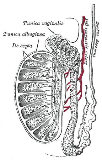
| The Tunica Albuginea is the fibrous covering of the testis. It is a dense membrane, of a bluish-white color, composed of bundles of white fibrous tissue which interlace in every direction. It is covered by the tunica vaginalis, except at the points of attachment of the epididymis to the testis, and along its posterior border, where the spermatic vessels enter the gland. It is applied to the tunica vasculosa over the glandular substance of the testis, and, at its posterior border, is reflected into the interior of the gland, forming an incomplete vertical septum, called the mediastinum testis (corpus Highmori). |
| The mediastinum testis extends from the upper to near the lower extremity of the gland, and is wider above than below. From its front and sides numerous imperfect septa (trabeculæ) are given off, which radiate toward the surface of the organ, and are attached to the tunica albuginea. They divide the interior of the organ into a number of incomplete spaces which are somewhat cone-shaped, being broad at their bases at the surface of the gland, and becoming narrower as they converge to the mediastinum. The mediastinum supports the vessels and duct of the testis in their passage to and from the substance of the gland. |
| The Tunica Vasculosa is the vascular layer of the testis, consisting of a plexus of bloodvessels, held together by delicate areolar tissue. It clothes the inner surface of the tunica albuginea and the different septa in the interior of the gland, and therefore forms an internal investment to all the spaces of which the gland is composed. |
 |
| FIG. 1149– Vertical section of the testis, to show the arrangement of the ducts. |
| |
| |
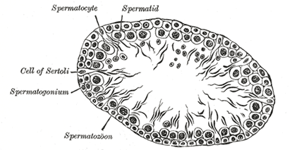
| Structure.—The glandular structure of the testis consists of numerous lobules. Their number, in a single testis, is estimated by Berres at 250, and by Krause at 400. They differ in size according to their position, those in the middle of the gland being larger and longer. The lobules (Fig. 1149) are conical in shape, the base being directed toward the circumference of the organ, the apex toward the mediastinum. Each lobule is contained in one of the intervals between the fibrous septa which extend between the mediastinum testis and the tunica albuginea, and consists of from one to three, or more, minute convoluted tubes, the tubuli seminiferi. The tubules may be separately unravelled, by careful dissection under water, and may be seen to commence either by free cecal ends or by anastomotic loops. They are supported by loose connective tissue which contains here and there groups of “interstitial cells” containing yellow pigment granules. The total number of tubules is estimated by Lauth at 840, and the average length of each is 70 to 80 cm. Their diameter varies from 0.12 to 0.3 mm. The tubules are pale in color in early life, but in old age they acquire a deep yellow tinge from containing much fatty matter. Each tubule consists of a basement layer formed of laminated connective tissue containing numerous elastic fibers with flattened cells between the layers and covered externally by a layer of flattened epithelioid cells. Within the basement membrane are epithelial cells arranged in several irregular layers, which are not always clearly separated, but which may be arranged in three different groups (Fig. 1150). Among these cells may be seen the spermatozoa in different stages of development. (1) Lining the basement membrane and forming the outer zone is a layer of cubical cells, with small nuclei; some of these enlarge to become spermatogonia. The nuclei of some of the spermatogonia may be seen to be in process of indirect division (karyokineses, page 37), and in consequence of this daughter cells are formed, which constitute the second zone. (2) Within this first layer is to be seen a number of larger polyhedral cells, with clear nuclei, arranged in two or three layers; these are the intermediate cells or spermatocytes. Most of these cells are in a condition of karyokinetic division, and the cells which result from this division form those of the next layer, the spermatoblasts or spermatids. (3) The third layer of cells consists of the spermatoblasts or spermatids, and each of these, without further subdivision, becomes a spermatozoön. The spermatids are small polyhedral cells, the nucleus of each of which contains half the usual number of chromosomes. In addition to these three layers of cells others are seen, which are termed the supporting cells (cells of Sertoli). They are elongated and columnar, and project inward from the basement membrane toward the lumen of the tube. As development of the spermatozoa proceeds the latter group themselves around the inner extremities of the supporting cells. The nuclear portion of the spermatid, which is partly imbedded in the supporting cell, is differentiated to form the head of the spermatozoön, while part of the cell protoplasm forms the middle piece and the tail is produced by an outgrowth from the double centriole of the cell. Ultimately the heads are liberated and the spermatozoa are set free. The structure of the spermatozoa is described on pages 42, 43. |
| In the apices of the lobules, the tubules become less convoluted, assume a nearly straight course, and unite together to form from twenty to thirty larger ducts, of about 0.5 mm. in diameter, and these, from their straight course, are called tubuli recti (Fig. 1149). |
 |
| FIG. 1150– Transverse section of a tubule of the testis of a rat. X 250. |
| |
| The tubuli recti enter the fibrous tissue of the mediastinum, and pass upward and backward, forming, in their ascent, a close net-work of anastomosing tubes which are merely channels in the fibrous stroma, lined by flattened epithelium, and having no proper walls; this constitutes the rete testis. At the upper end of the mediastinum, the vessels of the rete testis terminate in from twelve to fifteen or twenty ducts, the ductuli efferentes; they perforate the tunica albuginea, and carry the seminal fluid from the testis to the epididymis. Their course is at first straight; they then become enlarged, and exceedingly convoluted, and form a series of conical masses, the coni vasculosi, which together constitute the head of the epididymis. Each cone consists of a single convoluted duct, from 15 to 20 cm. in length, the diameter of which gradually decreases from the testis to the epididymis. Opposite the bases of the cones the efferent vessels open at narrow intervals into a single duct, which constitutes, by its complex convolutions, the body and tail of the epididymis. When the convolutions of this tube are unravelled, it measures upward of 6 meters in length; it increases in diameter and thickness as it approaches the ductus deferens. The convolutions are held together by fine areolar tissue, and by bands of fibrous tissue. |
 |
| FIG. 1151– Section of epididymis of guinea-pig. X 255. |
| |
| The tubuli recti have very thin walls; like the channels of the rete testis they are lined by a single layer of flattened epithelium. The ductuli efferentes and the tube of the epididymis have walls of considerable thickness, on account of the presence in them of muscular tissue, which is principally arranged in a circular manner. These tubes are lined by columnar ciliated epithelium (Fig. 1151). |
| |
| Peculiarities.—The testis, developed in the lumbar region, may be arrested or delayed in its transit to the scrotum (cryptorchism). It may be retained in the abdomen; or it may be arrested at the abdominal inguinal ring, or in the inguinal canal; or it may just pass out of the subcutaneous inguinal ring without finding its way to the bottom of the scrotum. When retained in the abdomen it gives rise to no symptoms, other than the absence of the testis from the scrotum; but when it is retained in the inguinal canal it is subjected to pressure and may become inflamed and painful. The retained testis is probably functionally useless; so that a man in whom both testes are retained (anorchism) is sterile, though he may not be impotent. The absence of one testis is termed monorchism. When a testis is retained in the inguinal canal it is often complicated with a congenital hernia, the funicular process of the peritoneum not being obliterated. In addition to the cases above described, where there is some arrest in the descent of the testis, this organ may descend through the inguinal canal, but may miss the scrotum and assume some abnormal position. The most common form is where the testis, emerging at the subcutaneous inguinal ring, slips down between the scrotum and thigh and comes to rest in the perineum. This is known as perineal ectopia testis. With each variety of abnormality in the position of the testis, it is very common to find concurrently a congenital hernia, or, if a hernia be not actually present, the funicular process is usually patent, and almost invariably so if the testis is in the inguinal canal. |
| The testis, finally reaching the scrotum, may occupy an abnormal position in it. It may be inverted, so that its posterior or attached border is directed forward and the tunica vaginalis is situated behind. |
| Fluid collections of a serous character are very frequently found in the scrotum. To these the term hydrocele is applied. The most common form is the ordinary vaginal hydrocele, in which the fluid is contained in the sac of the tunica vaginalis, which is separated, in its normal condition, from the peritoneal cavity by the whole extent of the inguinal canal. In another form, the congenital hydrocele, the fluid is in the sac of the tunica vaginalis, but this cavity communicates with the general peritoneal cavity, its tubular process remaining pervious. A third variety known as an infantile hydrocele, occurs in those cases where the tubular process becomes obliterated only at its upper part, at or near the abdominal inguinal ring. It resembles the vaginal hydrocele, except as regards its shape, the collection of fluid extending up the cord into the inguinal canal. Fourthly, the funicular process may become obliterated both at the abdominal inguinal ring and above the epididymis, leaving a central unobliterated portion, which may become distended with fluid, giving rise to a condition known as the encysted hydrocele of the cord. |


