 |
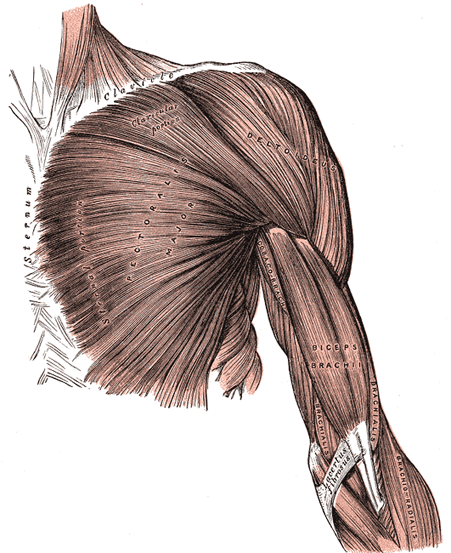
| FIG. 410– —Superficial muscles of the chest and front of the arm. |
|
| The muscles of the anterior and lateral thoracic regions are: |
| Pectoralis major. | |
Subclavius. |
| Pectoralis minor. | |
Serratus anterior. |
|
| |
| Superficial Fascia.—The superficial fascia of the anterior thoracic region is continuous with that of the neck and upper extremity above, and of the abdomen below. It encloses the mamma and gives off numerous septa which pass into the gland, supporting its various lobes. From the fascia over the front of the mamma, fibrous processes pass forward to the integument and papilla; these were called by Sir A. Cooper the ligamenta suspensoria. |
| |
| Pectoral Fascia.—The pectoral fascia is a thin lamina, covering the surface of the Pectoralis major, and sending numerous prolongations between its fasciculi: it is attached, in the middle line, to the front of the sternum; above, to the clavicle; laterally and below it is continuous with the fascia of the shoulder, axilla, and thorax. It is very thin over the upper part of the Pectoralis major, but thicker in the interval between it and the Latissimus dorsi, where it closes in the axillary space and forms the axillary fascia; it divides at the lateral margin of the Latissimus dorsi into two layers, one of which passes in front of, and the other behind it; these proceed as far as the spinous processes of the thoracic vertebræ, to which they are attached. As the fascia leaves the lower edge of the Pectoralis major to cross the floor of the axilla it sends a layer upward under cover of the muscle; this lamina splits to envelop the Pectoralis minor, at the upper edge of which it is continuous with the coracoclavicular fascia. The hollow of the armpit, seen when the arm is abducted, is produced mainly by the traction of this fascia on the axillary floor, and hence the lamina is sometimes named the suspensory ligament of the axilla. At the lower part of the thoracic region the deep fascia is well-developed, and is continuous with the fibrous sheaths of the Recti abdominis. |
| |
| The Pectoralis major (Fig. 410) is a thick, fan-shaped muscle, situated at the upper and forepart of the chest. It arises from the anterior surface of the sternal half of the clavicle; from half the breadth of the anterior surface of the sternum, as low down as the attachment of the cartilage of the sixth or seventh rib; from the cartilages of all the true ribs, with the exception, frequently, of the first or seventh, or both, and from the aponeurosis of the Obliquus externus abdominis. From this extensive origin the fibers converge toward their insertion; those arising from the clavicle pass obliquely downward and lateralward, and are usually separated from the rest by a slight interval; those from the lower part of the sternum, and the cartilages of the lower true ribs, run upward and lateralward; while the middle fibers pass horizontally. They all end in a flat tendon, about 5 cm. broad, which is inserted into the crest of the greater tubercle of the humerus. This tendon consists of two laminæ, placed one in front of the other, and usually blended together below. The anterior lamina, the thicker, receives the clavicular and the uppermost sternal fibers; they are inserted in the same order as that in which they arise: that is to say, the most lateral of the clavicular fibers are inserted at the upper part of the anterior lamina; the uppermost sternal fibers pass down to the lower part of the lamina which extends as low as the tendon of the Deltoideus and joins with it. The posterior lamina of the tendon receives the attachment of the greater part of the sternal portion and the deep fibers, i. e., those from the costal cartilages. These deep fibers, and particularly those from the lower costal cartilages, ascend the higher, turning backward successively behind the superficial and upper ones, so that the tendon appears to be twisted. The posterior lamina reaches higher on the humerus than the anterior one, and from it an expansion is given off which covers the intertubercular groove and blends with the capsule of the shoulder-joint. From the deepest fibers of this lamina at its insertion an expansion is given off which lines the intertubercular groove, while from the lower border of the tendon a third expansion passes downward to the fascia of the arm. |
| |
| Variations.—The more frequent variations are greater or less extent of attachment to the ribs and sternum, varying size of the abdominal part or its absence, greater or less extent of separation of sternocostal and clavicular parts, fusion of clavicular part with deltoid, decussation in front of the sternum. Deficiency or absence of the sternocostal part is not uncommon. Absence of the clavicular part is less frequent. Rarely the whole muscle is wanting. |
| Costocoracoideus is a muscular band occasionally found arising from the ribs or aponeurosis of the External oblique between the Pectoralis major and Latissimus dorsi and inserted into the coracoid process. |
| Chondro-epitrochlearis is a muscular slip occasionally found arising from the costal cartilages or from the aponeurosis of the External oblique below the Pectoralis major or from the Pectoralis major itself. The insertion is variable on the inner side of the arm to fascia, intermuscular septum or internal condyle. |
| Sternalis, in front of the sternal end of the Pectoralis major parallel to the margin of the sternum. It is supplied by the anterior thoracic nerves and is probably a misplaced part of the pectoralis. |
| |
| Coracoclavicular Fascia (fascia coracoclavicularis; costocoracoid membrane; clavipectoral fascia).—The coracoclavicular fascia is a strong fascia situated under cover of the clavicular portion of the Pectoralis major. It occupies the interval between the Pectoralis minor and Subclavius, and protects the axillary vessels and nerves. Traced upward, it splits to enclose the Subclavius, and its two layers are attached to the clavicle, one in front of and the other behind the muscle; the latter layer fuses with the deep cervical fascia and with the sheath of the axillary vessels. Medially, it blends with the fascia covering the first two intercostal spaces, and is attached also to the first rib medial to the origin of the Subclavius. Laterally, it is very thick and dense, and is attached to the coracoid process. The portion extending from the first rib to the coracoid process is often whiter and denser than the rest, and is sometimes called the costocoracoid ligament. Below this it is thin, and at the upper border of the Pectoralis minor it splits into two layers to invest the muscle; from the lower border of the Pectoralis minor it is continued downward to join the axillary fascia, and lateralward to join the fascia over the short head of the Biceps brachii. The coracoclavicular fascia is pierced by the cephalic vein, thoracoacromial artery and vein, and external anterior thoracic nerve. |
| The Pectoralis minor (Fig. 411) is a thin, triangular muscle, situated at the upper part of the thorax, beneath the Pectoralis major. It arises from the upper margins and outer surfaces of the third, fourth, and fifth ribs, near their cartilage and from the aponeuroses covering the Intercostalis; the fibers pass upward and lateralward and converge to form a flat tendon, which is inserted into the medial border and upper surface of the coracoid process of the scapula. |
| |
| Variations.—Origin from second, third and fourth or fifth ribs. The tendon of insertion may extend over the coracoid process to the greater tubercle. May be split into several parts. Absence rare. |
| Pectoralis minimus, first rib-cartilage to coracoid process. Rare. |
 |
| FIG. 411– Deep muscles of the chest and front of the arm, with the boundaries of the axilla. |
| |
| The Subclavius (Fig. 411) is a small triangular muscle, placed between the clavicle and the first rib. It arises by a short, thick tendon from the first rib and its cartilage at their junction, in front of the costoclavicular ligament; the fleshy fibers proceed obliquely upward and lateralward, to be inserted into the groove on the under surface of the clavicle between the costoclavicular and conoid ligaments. |
| |
| Variations.—Insertion into coracoid process instead of clavicle or into both clavicle and coracoid process. Sternoscapular fasciculus to the upper border of scapula. Sternoclavicularis from manubrium to clavicle between Pectoralis major and coracoclavicular fascia. |
| The Serratus anterior (Serratus magnus) (Fig. 411) is a thin muscular sheet, situated between the ribs and the scapula at the upper and lateral part of the chest. It arises by fleshy digitations from the outer surfaces and superior borders of the upper eight or nine ribs, and from the aponeuroses covering the intervening Intercostales. Each digitation (except the first) arises from the corresponding rib; the first springs from the first and second ribs; and from the fascia covering the first intercostal space. From this extensive attachment the fibers pass backward, closely applied to the chest-wall, and reach the vertebral border of the scapula, and are inserted into its ventral surface in the following manner. The first digitation is inserted into a triangular area on the ventral surface of the medial angle. The next two digitations spread out to form a thin, triangular sheet, the base of which is directed backward and is inserted into nearly the whole length of the ventral surface of the vertebral border. The lower five or six digitations converge to form a fan-shaped mass, the apex of which is inserted, by muscular and tendinous fibers, into a triangular impression on the ventral surface of the inferior angle. The lower four slips interdigitate at their origins with the upper five slips of the Obliquus externus abdominis. |
| |
| Variations.—Attachment to tenth rib. Absence of attachments to first rib, to one or more of the lower ribs. Division into three parts; absence or defect of middle part. Union with Levator scapulæ, External intercostals or External oblique. |
| |
| Nerves.—The Pectoralis major is supplied by the medial and lateral anterior thoracic nerves; through these nerves the muscle receives filaments from all the spinal nerves entering into the formation of the brachial plexus; the Pectoralis minor receives its fibers from the eighth cervical and first thoracic nerves through the medial anterior thoracic nerve. The Subclavius is suplied by a filament from the fifth and sixth cervical nerves; the Serratus anterior is supplied by the long thoracic, which is derived from the fifth, sixth, and seventh cervical nerves. |
| |
| Actions.—If the arm has been raised by the Deltoideus, the Pectoralis major will, conjointly with the Latissimus dorsi and Teres major, depress it to the side of the chest. If acting alone, it adducts and draws forward the arm, bringing it across the front of the chest, and at the same time rotates it inward. The Pectoralis minor depresses the point of the shoulder, drawing the scapula downward and medialward toward the thorax, and throwing the inferior angle backward. The Subclavius depresses the shoulder, carrying it downward and forward. When the arms are fixed, all three of these muscles act upon the ribs; drawing them upward and expanding the chest, and thus becoming very important agents in forced inspiration. The Serratus anterior, as a whole, carries the scapula forward, and at the same time raises the vertebral border of the bone. It is therefore concerned in the action of pushing. Its lower and stronger fibers move forward the lower angle and assist the Trapezius in rotating the bone at the sternoclavicular joint, and thus assist this muscle in raising the acromion and supporting weights upon the shoulder. It is also an assistant to the Deltoideus in raising the arm, inasmuch as during the action of this latter muscle it fixes the scapula and so steadies the glenoid cavity on which the head of the humerus rotates. After the Deltoideus has raised the arm to a right angle with the trunk, the Serratus anterior and the Trapezius, by rotating the scapula, raise the arm into an almost vertical position. It is possible that when the shoulders are fixed the lower fibers of the Serratus anterior may assist in raising and everting the ribs; but it is not the important inspiratory muscle it was formerly believed to be. |


