 |
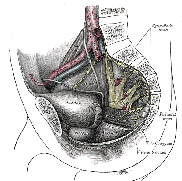
| FIG. 828– Plan of sacral and pudendal plexuses. |
|
(NN. Sacrales et Coccygeus) The anterior divisions of the sacral and coccygeal nerves (rami anteriores) form the sacral and pudendal plexuses. The anterior divisions of the upper four sacral nerves enter the pelvis through the anterior sacral foramina, that of the fifth between the sacrum and coccyx, while that of the coccygeal nerve curves forward below the rudimentary transverse process of the first piece of the coccyx. The first and second sacral nerves are large; the third, fourth, and fifth diminish progressively from above downward. Each receives a gray ramus communicans from the corresponding ganglion of the sympathetic trunk, while from the third and frequently from the second and the fourth sacral nerves, a white ramus communicans is given to the pelvic plexuses of the sympathetic. |
| |
| The Sacral Plexus (plexus sacralis) (Fig. 828).—The sacral plexus is formed by the lumbosacral trunk, the anterior division of the first, and portions of the anterior divisions of the second and third sacral nerves. |
| The lumbosacral trunk comprises the whole of the anterior division of the fifth and a part of that of the fourth lumbar nerve; it appears at the medial margin of the Psoas major and runs downward over the pelvic brim to join the first sacral nerve. The anterior division of the third sacral nerve divides into an upper and a lower branch, the former entering the sacral and the latter the pudendal plexus. |
| The nerves forming the sacral plexus converge toward the lower part of the greater sciatic foramen, and unite to form a flattened band, from the anterior and posterior surfaces of which several branches arise. The band itself is continued as the sciatic nerve. which splits on the back of the thigh into the tibial and common peroneal nerves; these two nerves sometimes arise separately from the plexus, and in all cases their independence can be shown by dissection. |
| |
| Relation.—The sacral plexus lies on the back of the pelvis between the Piriformis and the pelvic fascia (Fig. 829); in front of it are the hypogastric vessels, the ureter and the sigmoid colon. The superior gluteal vessels run between the lumbosacral trunk and the first sacral nerve, and the inferior gluteal vessels between the second and third sacral nerves. |
| All the nerves entering the plexus, with the exception of the third sacral, split into ventral and dorsal divisions, and the nerves arising from these are as follows: |
| | Ventral divisions. | Dorsal divisions. |
| Nerve to Quadratus femoris and Gemellus inferior | 4, 5 L, 1 S. | |
| Nerve to Obturator internus and Gemellus superior | 5 L, 1, 2 S. | |
| Nerve to Piriformis………………………………………………… | (1) 2 S. |
| Superior gluteal……………………………………………………. | 4, 5 L, 1 S. |
| Inferior gluteal……………………………………………………... | 5 L, 1, 2 S. |
| Posterior femoral cutaneous……………………… | 2, 3 S……… | 1, 2 S. |
| Sciatic | Tibial……………………………………… | 4, 5 L, 1, 2, 3 S. | |
Common peroneal………………………… | ……………… | 4, 5 L, 1, 2 S. | |
| The Nerve to the Quadratus Femoris and Gemellus Inferior arises from the ventral divisions of the fourth and fifth lumbar and first sacral nerves: it leaves the pelvis through the greater sciatic foramen, below the Piriformis, and runs down in front of the sciatic nerve, the Gemelli, and the tendon of the Obturator internus, and enters the anterior surfaces of the muscles; it gives an articular branch to the hip-joint. |
| |
| The Nerve to the Obturator Internus and Gemellus Superior arises from the ventral divisions of the fifth lumbar and first and second sacral nerves. It leaves the pelvis through the greater sciatic foramen below the Piriformis, and gives off the branch to the Gemellus superior, which enters the upper part of the posterior surface of the muscle. It then crosses the ischial spine, reënters the pelvis through the lesser sciatic foramen, and pierces the pelvic surface of the Obturator internus. |
| The Nerve to the Piriformis arises from the dorsal division of the second sacral nerve, or the dorsal divisions of the first and second sacral nerves, and enters the anterior surface of the muscle; this nerve may be double. |
 |
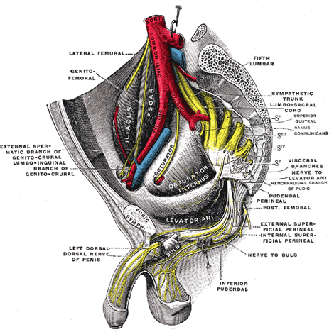
| FIG. 829– Dissection of side wall of pelvis showing sacral and pudendal plexuses. (Testut.) |
| |
| The Superior Gluteal Nerve (n. glutæus superior) arises from the dorsal divisions of the fourth and fifth lumbar and first sacral nerves: it leaves the pelvis through the greater sciatic foramen above the Piriformis, accompanied by the superior gluteal vessels, and divides into a superior and an inferior branch. The superior branch accompanies the upper branch of the deep division of the superior gluteal artery and ends in the Glutæus minimus. The inferior branch runs with the lower branch of the deep division of the superior gluteal artery across the Glutæus minimus; it gives filaments to the Glutæi medius and minimus, and ends in the Tensor fasciæ latæ. |
| The Inferior Gluteal Nerve (n. glutæus inferior) arises from the dorsal divisions of the fifth lumbar and first and second sacral nerves: it leaves the pelvis through the greater sciatic foramen, below the Piriformis, and divides into branches which enter the deep surface of the Glutæus maximus. |
| The Posterior Femoral Cutaneous Nerve (n. cutaneus femoralis posterior; small sciatic nerve) is distributed to the skin of the perineum and posterior surface of the thigh and leg. It arises partly from the dorsal divisions of the first and second, and from the ventral divisions of the second and third sacral nerves, and issues from the pelvis through the greater sciatic foramen below the Piriformis. It then descends beneath the Glutæus maximus with the inferior gluteal artery, and runs down the back of the thigh beneath the fascia lata, and over the long head of the Biceps femoris to the back of the knee; here it pierces the deep fascia and accompanies the small saphenous vein to about the middle of the back of the leg, its terminal twigs communicating with the sural nerve. |
| Its branches are all cutaneous, and are distributed to the gluteal region, the perineum, and the back of the thigh and leg. |
| The gluteal branches (nn. clunium inferiores), three or four in number, turn upward around the lower border of the Glutæus maximus, and supply the skin covering the lower and lateral part of that muscle. |
| The perineal branches (rami perineales) are distributed to the skin at the upper and medial side of the thigh. One long perineal branch, inferior pudendal (long scrotal nerve), curves forward below and in front of the ischial tuberosity, pierces the fascia lata, and runs forward beneath the superficial fascia of the perineum to the skin of the scrotum in the male, and of the labium majus in the female. It communicates with the inferior hemorrhoidal and posterior scrotal nerves. |
| The branches to the back of the thigh and leg consist of numerous filaments derived from both sides of the nerve, and distributed to the skin covering the back and medial side of the thigh, the popliteal fossa, and the upper part of the back of the leg (Fig. 830). |
| The Sciatic (n. ischiadicus; great sciatic nerve) (Fig. 832) supplies nearly the whole of the skin of the leg, the muscles of the back of the thigh, and those of the leg and foot. It is the largest nerve in the body, measuring 2 cm. in breadth, and is the continuation of the flattened band of the sacral plexus. It passes out of the pelvis through the greater sciatic foramen, below the Piriformis muscle. It descends between the greater trochanter of the femur and the tuberosity of the ischium, and along the back of the thigh to about its lower third, where it divides into two large branches, the tibial and common peroneal nerves. This division may take place at any point between the sacral plexus and the lower third of the thigh. When it occurs at the plexus, the common peroneal nerve usually pierces the Piriformis. |
| In the upper part of its course the nerve rests upon the posterior surface of the ischium, the nerve to the Quadratus femoris, the Obturator internus and Gemelli, and the Quadratus femoris; it is accompanied by the posterior femoral cutaneous nerve and the inferior gluteal artery, and is covered by the Glutæus maximus. Lower down, it lies upon the Adductor magnus, and is crossed obliquely by the long head of the Biceps femoris. |
| The nerve gives off articular and muscular branches. |
| The articular branches (rami articulares) arise from the upper part of the nerve and supply the hip-joint, perforating the posterior part of its capsule; they are sometimes derived from the sacral plexus. |
| The muscular branches (rami musculares) are distributed to the Biceps femoris, Semitendinosus, Semimembranosus, and Adductor magnus. The nerve to the short head of the Biceps femoris comes from the common peroneal part of the sciatic, while the other muscular branches arise from the tibial portion, as may be seen in those cases where there is a high division of the sciatic nerve. |
| The Tibial Nerve (n. tibialis; internal popliteal nerve) (Fig. 832) the larger of the two terminal branches of the sciatic, arises from the anterior branches of the fourth and fifth lumbar and first, second, and third sacral nerves. It descends along the back of the thigh and through the middle of the popliteal fossa, to the lower part of the Popliteus muscle, where it passes with the popliteal artery beneath the arch of the Soleus. It then runs along the back of the leg with the posterior tibial vessels to the interval between the medial malleolus and the heel, where it divides beneath the laciniate ligament into the medial and lateral plantar nerves. In the thigh it is overlapped by the hamstring muscles above, and then becomes more superficial, and lies lateral to, and some distance from, the popliteal vessels;opposite the knee-joint, it is in close relation with these vessels, and crosses to the medial side of the artery. In the leg it is covered in the upper part of its course by the muscles of the calf; lower down by the skin, the superficial and deep fasciæ. It is placed on the deep muscles, and lies at first to the medial side of the posterior tibial artery, but soon crosses that vessel and descends on its lateral side as far as the ankle. In the lower third of the leg it runs parallel with the medial margin of the tendo calcaneus. |
 |
| FIG. 830– Cutaneous nerves of right lower extremity. Posterior view. 137 |
| |
 |
| FIG. 831– Diagram of the segmental distribution of the cutaneous nerves of the right lower extremity. Posterior view. |
| |
| The branches of this nerve are: articular, muscular, medial sural cutaneous, medial calcaneal, medial and lateral plantar. |
| Articular branches (rami articulares), usually three in number, supply the knee-joint; two of these accompany the superior and inferior medial genicular arteries; and a third, the middle genicular artery. Just above the bifurcation of the nerve an articular branch is given off to the ankle-joint. |
| Muscular branches (rami musculares), four or five in number, arise from the nerve as it lies between the two heads of the Gastrocnemius muscle; they supply that muscle, and the Plantaris, Soleus, and Popliteus. The branch for the Popliteus turns around the lower border and is distributed to the deep surface of the muscle. Lower down, muscular branches arise separately or by a common trunk and supply the Soleus, Tibialis posterior, Flexor digitorum longus, and Flexor hallucis longus; the branch to the last muscle accompanies the peroneal artery; that to the Soleus enters the deep surface of the muscle. |
| The medial sural cutaneous nerve (n. cutaneus suræ medialis; n. communicans tibialis) descends between the two heads of the Gastrocnemius, and, about the middle of the back of the leg, pierces the deep fascia, and unites with the anastomotic ramus of the common peroneal to form the sural nerve (Fig. 830). |
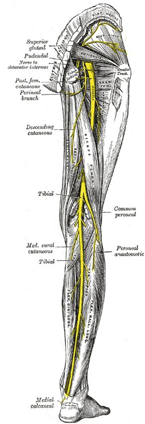 |
| FIG. 832– Nerves of the right lower extremity Posterior view. |
| |
| The sural nerve (n. suralis; short saphenous nerve), formed by the junction of the medial sural cutaneous with the peroneal anastomotic branch, passes downward near the lateral margin of the tendo calcaneus, lying close to the small saphenous vein, to the interval between the lateral malleolus and the calcaneus. It runs forward below the lateral malleolus, and is continued as the lateral dorsal cutaneous nerve along the lateral side of the foot and little toe, communicating on the dorsum of the foot with the intermediate dorsal cutaneous nerve, a branch of the superficial peroneal. In the leg, its branches communicate with those of the posterior femoral cutaneous. |
| The medial calcaneal branches (rami calcanei mediales; internal calcaneal branches) perforate the laciniate ligament, and supply the skin of the heel and medial side of the sole of the foot. |
| The medial plantar nerve (n. plantaris medialis; internal plantar nerve) (Fig. 833), the larger of the two terminal divisions of the tibial nerve, accompanies the medial plantar artery. From its origin under the laciniate ligament it passes under cover of the Abductor hallucis, and, appearing between this muscle and the Flexor digitorum brevis, gives off a proper digital plantar nerve and finally divides opposite the bases of the metatarsal bones into three common digital plantar nerves. |
| |
| BRANCHES.—The branches of the medial plantar nerve are: (1) cutaneous, (2) muscular, (3) articular, (4) a proper digital nerve to the medial side of the great toe, and (5) three common digital nerves. |
| The cutaneous branches pierce the plantar aponeurosis between the Abductor hallucis and the Flexor digitorum brevis and are distributed to the skin of the sole of the foot. |
| The muscular branches supply the Abductor hallucis, the Flexor digitorum brevis, the Flexor hallucis brevis, and the first Lumbricalis; those for the Abductor hallucis and Flexor digitorum brevis arise from the trunk of the nerve near its origin and enter the deep surfaces of the muscles; the branch of the Flexor hallucis brevis springs from the proper digital nerve to the medial side of the great toe, and that for the first Lumbricalis from the first common digital nerve. |
| The articular branches supply the articulations of the tarsus and metatarsus. |
| The proper digital nerve of the great toe (nn. digitales plantares proprii; plantar digital branches) supplies the Flexor hallucis brevis and the skin on the medial side of the great toe. |
| The three common digital nerves (nn. digitales plantares communes) pass between the divisions of the plantar aponeurosis, and each splits into two proper digital nerves—those of the first common digital nerve supply the adjacent sides of the great and second toes; those of the second, the adjacent sides of the second and third toes; and those of the third, the adjacent sides of the third and fourth toes. The third common digital nerve receives a communicating branch from the lateral plantar nerve; the first gives a twig to the first Lumbricalis. Each proper digital nerve gives off cutaneous and articular filaments; and opposite the last phalanx sends upward a dorsal branch, which supplies the structures around the nail, the continuation of the nerve being distributed to the ball of the toe. It will be observed that these digital nerves are similar in their distribution to those of the median nerve in the hand. |
| The Lateral Plantar Nerve (n. plantaris lateralis; external plantar nerve) (Fig. 833) supplies the skin of the fifth toe and lateral half of the fourth, as well as most of the deep muscles, its distribution being similar to that of the ulnar nerve in the hand. It passes obliquely forward with the lateral plantar artery to the lateral side of the foot, lying between the Flexor digitorum brevis and Quadratus plantæ and, in the interval between the former muscle and the Abductor digiti quinti, divides into a superficial and a deep branch. Before its division, it supplies the Quadratus plantæ and Abductor digiti quinti. |
| The superficial branch (ramus superficialis) splits into a proper and a common digital nerve; the proper digital nerve supplies the lateral side of the little toe, the Flexor digiti quinti brevis, and the two Interossei of the fourth intermetatarsal space; the common digital nerve communicates with the third common digital branch of the medial plantar nerve and divides into two proper digital nerves which supply the adjoining sides of the fourth and fifth toes. |
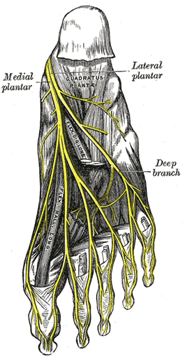 |
| FIG. 833– The plantar nerves. |
| |
 |
| FIG. 834– Diagram of the segmental distribution of the cutaneous nerves of the sole of the foot. |
| |
| The deep branch (ramus profundus; muscular branch) accompanies the lateral plantar artery on the deep surface of the tendons of the Flexor muscles and the Adductor hallucis, and supplies all the Interossei (except those in the fourth metatarsal space), the second, third, and fourth Lumbricales, and the Adductor hallucis. |
| The Common Peroneal Nerve (n. peronæus communis; external popliteal nerve; peroneal nerve) (Fig. 832), about one-half the size of the tibial, is derived from the dorsal branches of the fourth and fifth lumbar and the first and second sacral nerves. It descends obliquely along the lateral side of the popliteal fossa to the head of the fibula, close to the medial margin of the Biceps femoris muscle. It lies between the tendon of the Biceps femoris and lateral head of the Gastrocnemius muscle, winds around the neck of the fibula, between the Peronæus longus and the bone, and divides beneath the muscle into the superficial and deep peroneal nerves. Previous to its division it gives off articular and lateral sural cutaneous nerves. |
| The articular branches (rami articulares) are three in number; two of these accompany the superior and inferior lateral genicular arteries to the knee; the upper one occasionally arises from the trunk of the sciatic nerve. The third (recurrent) articular nerve is given off at the point of division of the common peroneal nerve; it ascends with the anterior recurrent tibial artery through the Tibialis anterior to the front of the knee. |
| The lateral sural cutaneous nerve (n. cutaneus suræ lateralis; lateral cutaneous branch) supplies the skin on the posterior and lateral surfaces of the leg; one branch, the peroneal anastomotic (n. communicans fibularis), arises near the head of the fibula, crosses the lateral head of the Gastrocnemius to the middle of the leg, and joins with the medial sural cutaneous to form the sural nerve. The peroneal anastomotic is occasionally continued down as a separate branch as far as the heel. |
| The Deep Peroneal Nerve (n. peronæus profundus; anterior tibial nerve) (Fig. 827) begins at the bifurcation of the common peroneal nerve, between the fibula and upper part of the Peronæus longus, passes obliquely forward beneath the Extensor digitorum longus to the front of the interosseous membrane, and comes into relation with the anterior tibial artery above the middle of the leg; it then descends with the artery to the front of the ankle-joint, where it divides into a lateral and a medial terminal branch. It lies at first on the lateral side of the anterior tibial artery, then in front of it, and again on its lateral side at the ankle-joint. |
| In the leg, the deep peroneal nerve supplies muscular branches to the Tibialis anterior, Extensor digitorum longus, Peronæus tertius, and Extensor hallucis prop ius, and an articular branch to the ankle-joint. |
| The lateral terminal branch (external or tarsal branch) passes across the tarsus, beneath the Extensor digitorum brevis, and, having become enlarged like the dorsal interosseous nerve at the wrist, supplies the Extensor digitorumbrevis. From the enlargement three minute interosseous branches are given off, which supply the tarsal joints and the metatarsophalangeal joints of the second, third, and fourth toes. The first of these sends a filament to the second Interosseus dorsalis muscle. |
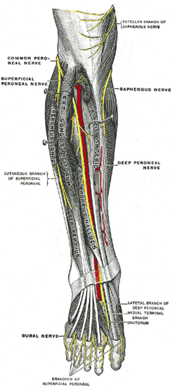 |
| FIG. 835– Deep nerves of the front of the leg. (Testut.) |
| |
| The medial terminal branch (internal branch) accompanies the dorsalis pedis artery along the dorsum of the foot, and, at the first interosseous space, divides into two dorsal digital nerves (nn. digitales dorsales hallucis lateralis et digiti secundi medialis) which supply the adjacent sides of the great and second toes, communicating with the medial dorsal cutaneous branch of the superficial peroneal nerve. Before it divides it gives off to the first space an interosseous branch which supplies the metatarsophalangeal joint of the great toe and sends a filament to the first Interosseous dorsalis muscle. |
| The Superficial Peroneal Nerve (n. peronæus superficialis; musculocutaneous nerve) (Figs. 827, 835) supplies the Peronei longus and brevis and the skin over the greater part of the dorsum of the foot. It passes forward between the Peronæi and the Extensor digitorum longus, pierces the deep fascia at the lower third of the leg, and divides into a medial and an intermediate dorsal cutaneous nerve. In its course between the muscles, the nerve gives off muscular branches to the Peronæi longus and brevis, and cutaneous filaments to the integument of the lower part of the leg. |
 |
| FIG. 836– Nerves of the dorsum of the foot. (Testut.) |
| |
| The medial dorsal cutaneous nerve (n. cutaneus dorsalis medialis; internal dorsal cutaneous branch) passes in front of the ankle-joint, and divides into two dorsal digital branches, one of which supplies the medial side of the great toe, the other, the adjacent side of the second and third toes. It also supplies the integument of the medial side of the foot and ankle, and communicates with the saphenous nerve, and with the deep peroneal nerve (Fig. 825). |
| The intermediate dorsal cutaneous nerve (n. cutaneus dorsalis intermedius; external dorsal cutaneous branch), the smaller, passes along the lateral part of the dorsum of the foot, and divides into dorsal digital branches, which supply the contiguous sides of the third and fourth, and of the fourth and fifth toes. It also supplies the skin of the lateral side of the foot and ankle, and communicates with the sural nerve (Fig. 825). The branches of the superficial peroneal nerve supply the skin of the dorsal surfaces of all the toes excepting the lateral side of the little toe, and the adjoining sides of the great and second toes, the former being supplied by the lateral dorsal cutaneous nerve from the sural nerve, and the latter by the medial branch of the deep peroneal nerve. Frequently some of the lateral branches of the superficial peroneal are absent, and their places are then taken by branches of the sural nerve. |
| |
| The Pudendal Plexus (plexus pudendus) (Fig. 828).—The pudendal plexus is not sharply marked off from the sacral plexus, and as a consequence some of the branches which spring from it may arise in conjunction with those of the sacral plexus. It lies on the posterior wall of the pelvis, and is usually formed by branches from the anterior divisions of the second and third sacral nerves, the whole of the anterior divisions of the fourth and fifth sacral nerves, and the coccygeal nerve. |
| It gives off the following branches: |
| Perforating cutaneous… | 2, 3 S. |
| Pudendal……………… | 2, 3, 4 S. |
| Visceral……………… | 3, 4 S. |
| Muscular……………… | 4 S. |
| Anococcygeal………… | 4, 5 S. and Cocc. | |
 |
| FIG. 837– Sacral plexus of the right side. (Testut). |
| |
| The Perforating Cutaneous Nerve (n. clunium inferior medialis) usually arises from the posterior surface of the second and third sacral nerves. It pierces the lower part of the sacrotuberous ligament, and winding around the inferior border of the Glutæus maximus supplies the skin covering the medial and lower parts of that muscle. |
| The perforating cutaneous nerve may arise from the pudendal or it may be absent; in the latter case its place may be taken by a branch from the posterior femoral cutaneous nerve or by a branch from the third and fourth, or fourth and fifth, sacral nerves. |
| The Pudendal Nerve (n. pudendus; internal pudic nerve) derives its fibers from the ventral branches of the second, third, and fourth sacral nerves. It passes between the Piriformis and Coccygeus muscles and leaves the pelvis through the lower part of the greater sciatic foramen. It then crosses the spine of the ischium, and reënters the pelvis through the lesser sciatic foramen. It accompanies the internal pudendal vessels upward and forward along the lateral wall of the ischiorectal fossa, being contained in a sheath of the obturator fascia termed Alcock’s canal, and divides into two terminal branches, viz., the perineal nerve, and the dorsal nerve of the penis or clitoris. Before its division it gives off the inferior hemorrhoidal nerve. |
| The inferior hemorrhoidal nerve (n. hæmorrhoidalis inferior) occasionally arises directly from the sacral plexus; it crosses the ischiorectal fossa, with the inferior hemorrhoidal vessels, toward the anal canal and the lower end of the rectum, and is distributed to the Sphincter ani externus and to the integument around the anus. Branches of this nerve communicate with the perineal branch of the posterior femoral cutaneous and with the posterior scrotal nerves at the forepart of the perineum. |
| The perineal nerve (n. perinei), the inferior and larger of the two terminal branches of the pudendal, is situated below the internal pudendal artery. It accompanies the perineal artery and divides into posterior scrotal (or labial) and muscular branches. |
| The posterior scrotal (or labial) branches (nn. scrotales (or labiales) posteriores; superficial peroneal nerves) are two in number, medial and lateral. They pierce the fascia of the urogenital diaphragm, and run forward along the lateral part of the urethral triangle in company with the posterior scrotal branches of the perineal artery; they are distributed to the skin of the scrotum and communicate with the perineal branch of the posterior femoral cutaneous nerve. These nerves supply the labium majus in the female. |
| The muscular branches are distributed to the Transversus perinæi superficialis, Bulbocavernous, Ischiocavernosus, and Constrictor urethræ. A branch, the nerve to the bulb, given off from the nerve to the Bulbocavernosus, pierces this muscle, and supplies the corpus cavernosum urethræ, ending in the mucous membrane of the urethra. |
| The dorsal nerve of the penis (n. dorsalis penis) is the deepest division of the pudendal nerve; it accompanies the internal pudendal artery along the ramus of the ischium; it then runs forward along the margin of the inferior ramus of the pubis, between the superior and inferior layers of the fascia of the urogenital diaphragm. Piercing the inferior layer it gives a branch to the corpus cavernosum penis, and passes forward, in company with the dorsal artery of the penis, between the layers of the suspensory ligament, on to the dorsum of the penis, and ends on the glans penis. In the female this nerve is very small, and supplies the clitoris (n. dorsalis clitoridis). |
| The Visceral Branches arise from the third and fourth, and sometimes from the second, sacral nerves, and are distributed to the bladder and rectum and, in the female, to the vagina; they communicate with the pelvic plexuses of the sympathetic. |
| The Muscular Branches are derived from the fourth sacral, and supply the Levator ani, Coccygeus, and Sphincter ani externus. The branches to the Levator ani and Coccygeus enter their pelvic surfaces; that to the Sphincter ani externus (perineal branch) reaches the ischiorectal fossa by piercing the Coccygeus or by passing between it and the Levator ani. Cutaneous filaments from this branch supply the skin between the anus and the coccyx. |
| |
| Anococcygeal Nerves (nn. anococcygei).—The fifth sacral nerve receives a communicating filament from the fourth, and unites with the coccygeal nerve to form the coccygeal plexus. From this plexus the anococcygeal nerves take origin; they consist of a few fine filaments which pierce the sacrotuberous ligament to supply the skin in the region of the coccyx. |


