6c. 5. The Tibia
 |
(Shin Bone) The tibia (Figs. 258, 259) is situated at the medial side of the leg, and, excepting the femur, is the longest bone of the skeleton. It is prismoid in form, expanded above, where it enters into the knee-joint, contracted in the lower third, and again enlarged but to a lesser extent below. In the male, its direction is vertical, and parallel with the bone of the opposite side; but in the female it has a slightly oblique direction downward and lateralward, to compensate for the greater obliquity of the femur. It has a body and two extremities. |
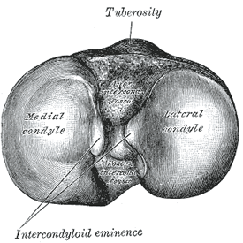
| The Upper Extremity (proximal extremity).—The upper extremity is large, and expanded into two eminences, the medial and lateral condyles. The superior articular surface presents two smooth articular facets (Fig. 257). The medial facet, oval in shape, is slightly concave from side to side, and from before backward. The lateral, nearly circular, is concave from side to side, but slightly convex from before backward, especially at its posterior part, where it is prolonged on to the posterior surface for a short distance. The central portions of these facets articulate with the condyles of the femur, while their peripheral portions support the menisci of the knee-joint, which here intervene between the two bones. Between the articular facets, but nearer the posterior than the anterior aspect of the bone, is the intercondyloid eminence (spine of tibia), surmounted on either side by a prominent tubercle, on to the sides of which the articular facets are prolonged; in front of and behind the intercondyloid eminence are rough depressions for the attachment of the anterior and posterior cruciate ligaments and the menisci. The anterior surfaces of the condyles are continuous with one another, forming a large somewhat flattened area; this area is triangular, broad above, and perforated by large vascular foramina; narrow below where it ends in a large oblong elevation, the tuberosity of the tibia, which gives attachment to the ligamentum patellæ; a bursa intervenes between the deep surface of the ligament and the part of the bone immediately above the tuberosity. Posteriorly, the condyles are separated from each other by a shallow depression, the posterior intercondyloid fossa, which gives attachment to part of the posterior cruciate ligament of the knee-joint. The medial condyle presents posteriorly a deep transverse groove, for the insertion of the tendon of the Semimembranosus. Its medial surface is convex, rough, and prominent; it gives attachment to the tibial collateral ligament. The lateral condyle presents posteriorly a flat articular facet, nearly circular in form, directed downward, backward, and lateralward, for articulation with the head of the fibula. Its lateral surface is convex, rough, and prominent in front: on it is an eminence, situated on a level with the upper border of the tuberosity and at the junction of its anterior and lateral surfaces, for the attachment of the iliotibial band. Just below this a part of the Extensor digitorum longus takes origin and a slip from the tendon of the Biceps femoris is inserted. |
| The Body or Shaft (corpus tibiæ).—The body has three borders and three surfaces. |
| Borders.—The anterior crest or border, the most prominent of the three, commences above at the tuberosity, and ends below at the anterior margin of the medial malleolus. It is sinuous and prominent in the upper two-thirds of its extent, but smooth and rounded below; it gives attachment to the deep fascia of the leg. |
| The medial border is smooth and rounded above and below, but more prominent in the center; it begins at the back part of the medial condyle, and ends at the posterior border of the medial malleolus; its upper part gives attachment to the tibial collateral ligament of the knee-joint to the extent of about 5 cm., and insertion to some fibers of the Popliteus; from its middle third some fibers of the Soleus and Flexor digitorum longus take origin. |
| The interosseous crest or lateral border is thin and prominent, especially its central part, and gives attachment to the interosseous membrane; it commences above in front of the fibular articular facet, and bifurcates below, to form the boundaries of a triangular rough surface, for the attachment of the interosseous ligament connecting the tibia and fibula. |
 |
| Surfaces.—The medial surface is smooth, convex, and broader above than below; its upper third, directed forward and medialward, is covered by the aponeurosis derived from the tendon of the Sartorius, and by the tendons of the Gracilis and Semitendinosus, all of which are inserted nearly as far forward as the anterior crest; in the rest of its extent it is subcutaneous. |
| The lateral surface is narrower than the medial; its upper two-thirds present a shallow groove for the origin of the Tibialis anterior; its lower third is smooth, convex, curves gradually forward to the anterior aspect of the bone, and is covered by the tendons of the Tibialis anterior, Extensor hallucis longus, and Extensor digitorum longus, arranged in this order from the medial side. |
 |
| The posterior surface (Fig. 259) presents, at its upper part, a prominent ridge, the popliteal line, which extends obliquely downward from the back part of the articular facet for the fibula to the medial border, at the junction of its upper and middle thirds; it marks the lower limit of the insertion of the Popliteus, serves for the attachment of the fascia covering this muscle, and gives origin to part of the Soleus, Flexor digitorum longus, and Tibialis posterior. The triangular area, above this line, gives insertion to the Popliteus. The middle third of the posterior surface is divided by a vertical ridge into two parts; the ridge begins at the popliteal line and is well-marked above, but indistinct below; the medial and broader portion gives origin to the Flexor digitorum longus, the lateral and narrower to part of the Tibialis posterior. The remaining part of the posterior surface is smooth and covered by the Tibialis posterior, Flexor digitorum longus, and Flexor hallucis longus. Immediately below the popliteal line is the nutrient foramen, which is large and directed obliquely downward. |
| The Lower Extremity (distal extremity).—The lower extremity, much smaller than the upper, presents five surfaces; it is prolonged downward on its medial side as a strong process, the medial malleolus. |
| Surfaces.—The inferior articular surface is quadrilateral, and smooth for articulation with the talus. It is concave from before backward, broader in front than behind, and traversed from before backward by a slight elevation, separating two depressions. It is continuous with that on the medial malleolus. |
 |
 |
| The anterior surface of the lower extremity is smooth and rounded above, and covered by the tendons of the Extensor muscles; its lower margin presents a rough transverse depression for the attachment of the articular capsule of the ankle-joint. |
| The posterior surface is traversed by a shallow groove directed obliquely downward and medialward, continuous with a similar groove on the posterior surface of the talus and serving for the passage of the tendon of the Flexor hallucis longus. |
| The lateral surface presents a triangular rough depression for the attachment of the inferior interosseous ligament connecting it with the fibula; the lower part of this depression is smooth, covered with cartilage in the fresh state, and articulates with the fibula. The surface is bounded by two prominent borders, continuous above with the interosseous crest; they afford attachment to the anterior and posterior ligaments of the lateral malleolus. |
| The medial surface is prolonged downward to form a strong pyramidal process, flattened from without inward—the medial malleolus. The medial surface of this process is convex and subcutaneous; its lateral or articular surface is smooth and slightly concave, and articulates with the talus; its anterior border is rough, for the attachment of the anterior fibers of the deltoid ligament of the ankle-joint; its posterior border presents a broad groove, the malleolar sulcus, directed obliquely downward and medialward, and occasionally double; this sulcus lodges the tendons of the Tibialis posterior and Flexor digitorum longus. The summit of the medial malleolus is marked by a rough depression behind, for the attachment of the deltoid ligament. |
| Structure.—The structure of the tibia is like that of the other long bones. The compact wall of the body is thickest at the junction of the middle and lower thirds of the bone. |
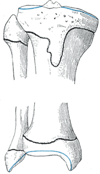
| Ossification.—The tibia is ossified from three centers (Figs. 260, 261): one for the body and one for either extremity. Ossification begins in the center of the body, about the seventh week of fetal life, and gradually extends toward the extremities. The center for the upper epiphysis appears before or shortly after birth; it is flattened in form, and has a thin tongue-shaped process in front, which forms the tuberosity (Fig. 260); that for the lower epiphysis appears in the second year. The lower epiphysis joins the body at about the eighteenth, and the upper one joins about the twentieth year. Two additional centers occasionally exist, one for the tongue-shaped process of the upper epiphysis, which forms the tuberosity, and one for the medial malleolus. |
